Radiomics: an aspect of COLOSSUS being incorporated into our research plan to bear down on the COLOSSUS-identified subtypes and further sub-stratify MSS RAS mutated colorectal cancer (CRC) to help develop new treatments and drugs.
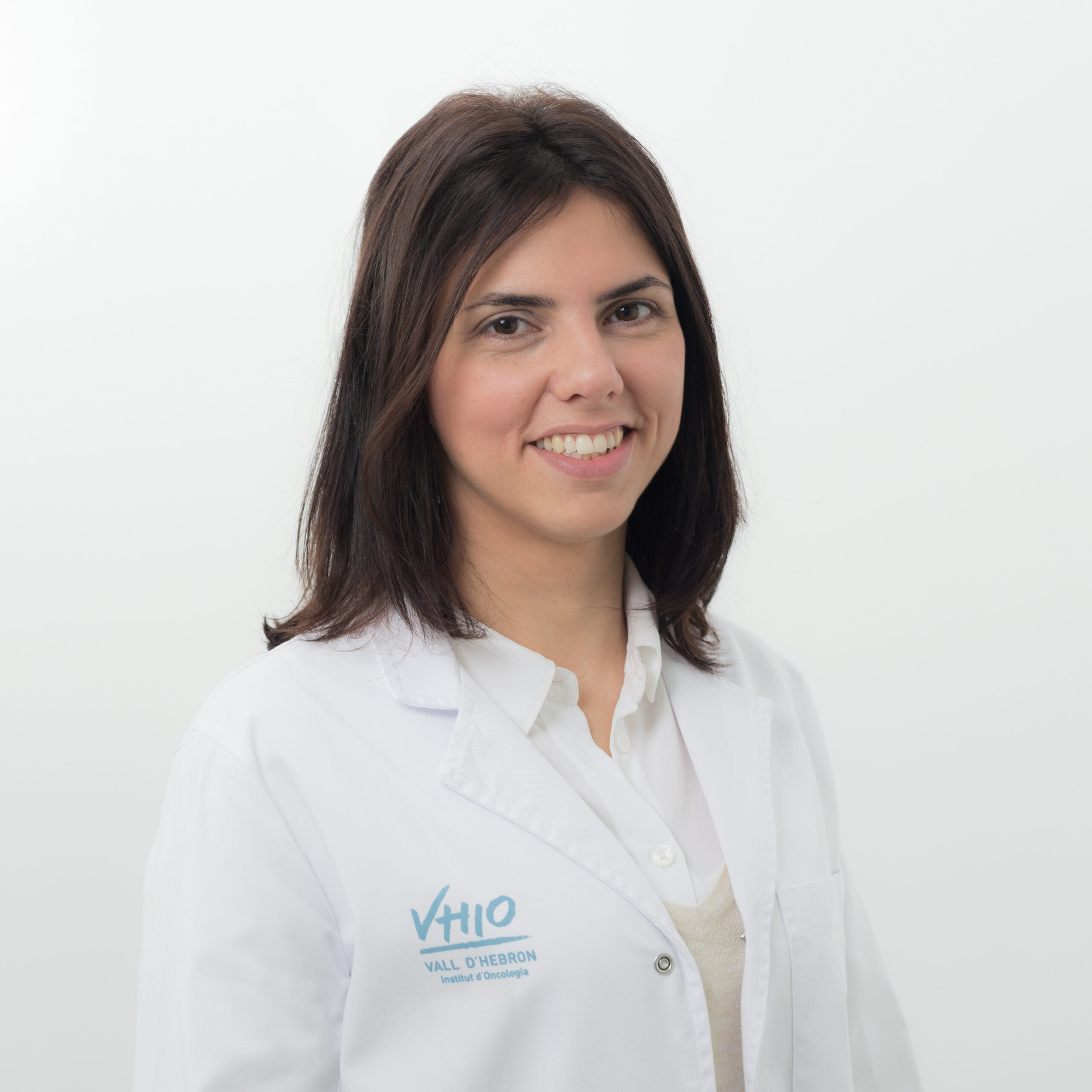
Dr Raquel Perez-Lopez is an experienced academic radiologist trained at the Royal Marsden Hospital and the Institute of Cancer Research in London (United Kingdom). Her PhD studies enabled her to identify the value of functional imaging (whole-body diffusion-weighted MRI) as a prognostic and response biomarker in prostate cancer which in turn led to the completion of the first prospective clinical trial in this context.
Dr Perez Lopez joined the Vall d’Hebron University Hospital, VHIO in the autumn of 2017 as Group Leader of VHIO’s newly established Radiomics Group, focused on the application of advanced imaging processing tools and integration of image data to other -omics. Her group seeks to improve cancer patients care by more effectively characterizing response to novel agents, identifying those patients who are most likely to benefit from these therapies, and further enforce VHIO’s translational research programmes in genomics, predictive science, and biomarkers of response and resistance. Raquel also works as a Consultant Radiologist in the Radiology Department at the Vall d’Hebron University Hospital. She is an experienced radiologist in oncological imaging with wide expertise in the application of imaging tools in clinical trials.
The Radiomics Group at VHIO
The Radiomics Group, led by Raquel Perez-Lopez, was established at Vall d’Hebron Institute of Oncology (VHIO) in October 2017. Their efforts centre on advancing precision imaging in personalized medicine, towards ultimately improving outcomes for cancer patients by applying mathematical methods to image processing and artificial intelligence in the biomedical field. Specifically, the Radiomics Group aims to: 1) integrate imaging, engineering and bioinformatics expertise towards the development and clinical qualification of quantitative imaging biomarkers for precision medicine, 2) integrate radiomics and genomics in translational studies towards the deeper understanding of tumour evolution and mechanisms of resistance to treatment, 3) develop and implement computational models for advanced image processing and 4) optimize and standardize imaging analysis pipelines.
The Radiomics Group recent work has resulted in several manuscripts in prestigious titles including Clinical Cancer Research and Radiology.
Radiomics
Medical imaging is widely used to perform non-invasive tumour assessment and treatment guidance in clinical practice. Advances in computational medical imaging analysis allow for large- and small-scale tumour features quantification using automatically extracted data characterization algorithms (radiomics). Radiomics signatures provide valuable information about the tumour molecular phenotype. Tumours present spatial and temporal biological heterogeneity, therefore, radiomics has great potential to guide personalized treatments because it can provide a comprehensive view of the entire tumour at several timepoints. The accurate characterization of the spatial-temporal landscape of tumour phenotype by non-invasive imaging assays may result in better tumour response assessment and aid the selection for effective therapy.
Overall, radiomics can be used to facilitate a deeper understanding of tumour biology, capture tumour heterogeneity and monitor tumour evolution and response to therapy.
The Radiomics Group’s role within the COLOSSUS project
Dr Raquel Perez-Lopez will lead and coordinate the medical imaging area of the COLOSSUS trial. She and the dedicated Radiomics Group’s members will be in charge of the: 1) computational analysis of the CT scans within the COLOSSUS trial, 2) application of AI-models for MSS RAS mutated CRC radiogenomics signatures development, 3) identification of radiomics characteristics that define the individual susceptibility of MSS RAS mutated subtypes to respond to treatment and 4) integration of radiomics with and other sources of information from the tumours such as genomics or transcriptomics, towards a better characterization of MSS RAS mutant CRC phenotypes and prediction of response to treatment.

